
| 规格 | 价格 | 库存 | 数量 |
|---|---|---|---|
| 1mg | ¥ 433.00 | 100 | |
| 5mg | ¥ 1628.00 | 100 | |
| 10mg | ¥ 2599.00 | 100 | |
| 25mg | ¥ 5959.00 | 100 |
| 别名 | LNS 8801 |
| 产品名 | G-1 |
| CAS号 | 881639-98-1 |
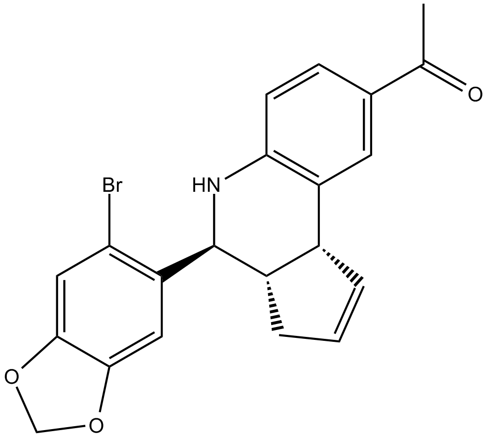
| 化学式 | C₂₁H₁₈BrNO₃ |
| 分子量 | 412.28 |
| 溶解度 | 1mg/ml in ethanol; 20mg/ml in DMSO; 30mg/ml in DMF |
| 靶点 | GPR30 agonist, potent and selective |
| 储存条件 | Store at -20°C |
| 用途 | 仅供科学研究使用!不能用于人体及动物治疗 |
| 纯度 | >99.3% |
靶点及简介 :GPR30 agonist, potent and selective
G-1 是一种 G蛋白偶联雌激素受体1(GPR30 或 GPER1)的非甾体抑制剂,其Ki值为11 nM,EC50值为2 nM。G-1 profoundly inhibited MCF-7 cell growth, potentially via p53 and p21 induction. Further, flow cytometry showed that G-1 blocked MCF-7 cell cycle progression at the G(1) phase.G-1 inhibits the production of lipopolysaccharide (LPS)-induced cytokines such as TNF-alpha and IL-6 in a dose-dependent manner in human primary macrophages and in a murine macrophage cell line. In vivo, G-1 is able to reduce the severity of disease in both active and passive EAE models of multiple sclerosis in SJL mice and that this effect is concomitant with a G-1-mediated decrease in proinflammatory cytokines, including IFN-gamma and IL-17, in immune cells harvested from these mice. The effect of G-1 appears indirect, as the GPR30 agonist did not directly influence IFN-gamma or IL-17 production by purified T cells. G-1 was able to induce both c-fos expression and proliferation in the ERalpha-negative/GPR30-positive SKBR3 breast cancer cells, the requirement for ERalpha expression in GPR30/EGFR signaling may depend on the specific cellular context of different tumor types.
体外研究
G-1是GPR30的非甾体,高亲和力和选择性激动剂,Ki为11 nM [1]。用G-1(10μM和100μM)处理48和72小时显着降低细胞增殖(P <0.001)。在72小时,计算G-1的IC 50值为20μM。用浓度为20μM的G-1处理A549细胞显示细胞凋亡显着增加,与其抗增殖作用一致(P <0.001)[2]。 G-1处理24小时后H295R细胞的细胞周期分析表明细胞周期停滞在G2期。 G-1的存在增加了Bax的表达,同时降低了Bcl-2 [3]。
体内研究
伤后14天的结果显示G-1组的Basso小鼠量表(BMS)评分显着高于其他组(P <0.05)。计数切片中caspase-3阳性细胞数,G-1组阳性细胞数少于其他组(P <0.05),两组间差异无统计学意义(P> 0.05) [1]。从治疗后第14天开始,G-1给药产生统计学上显着的肿瘤体积减少。与载体治疗的动物相比,用G-1治疗三周后收获的移植肿瘤显示肿瘤重量显着降低[3]。
细胞实验
在96孔板中用各种浓度(10nM,100nM,1μM,10μM和100μM)的G-1处理A549人肺癌细胞,并孵育48或72小时。温育后,将MTT溶液以0.5mg / mL的浓度添加到各孔中,并在37℃下孵育4小时。在此期间结束时,向每个孔中加入100μLDMSO溶剂。使用分光光度计[2]读取每个孔中溶液在570nm处的吸光度值[光密度(OD)]。
动物实验
在该研究中使用四周龄的nu / nu-Forkhead盒N1nu雌性小鼠。将悬浮在100μLPBS中的H295R细胞(6×10⁶)与30μL基质胶(4mg / mL)组合,并皮下注射到每只动物的肩部。在细胞注射后21天处理小鼠,此时肿瘤的平均体积达到约200mm 3。将动物随机分配用载体或G-1以2mg / kg /天的浓度处理。在以下方面评估荷瘤小鼠的药物耐受性:a)致死性毒性,即在对照小鼠中任何死亡之前发生的治疗小鼠中的任何死亡; b)体重减轻百分比= 100 - [(第x天的体重/第1天的体重)×100],其中x表示治疗期间的一天。注射细胞后42天,通过颈椎脱位处死动物[3]。
验证:
参考文献:
| 浓度/溶剂体积/质量 | 1 mg | 5 mg | 10 mg |
|---|---|---|---|
| 1 mM | 2.4255 mL | 12.1277 mL | 24.2554 mL |
| 5 mM | 0.4851 mL | 2.4255 mL | 4.8511 mL |
| 10 mM | 0.2426 mL | 1.2128 mL | 2.4255 mL |


长按屏幕识别二维码
打开手机扫描二维码